Two colors reveal defense signals in a glowing leaf
Bright leaf veins can obscure a plant’s response to infection, but comparing two colors of light helps bring that response into view.
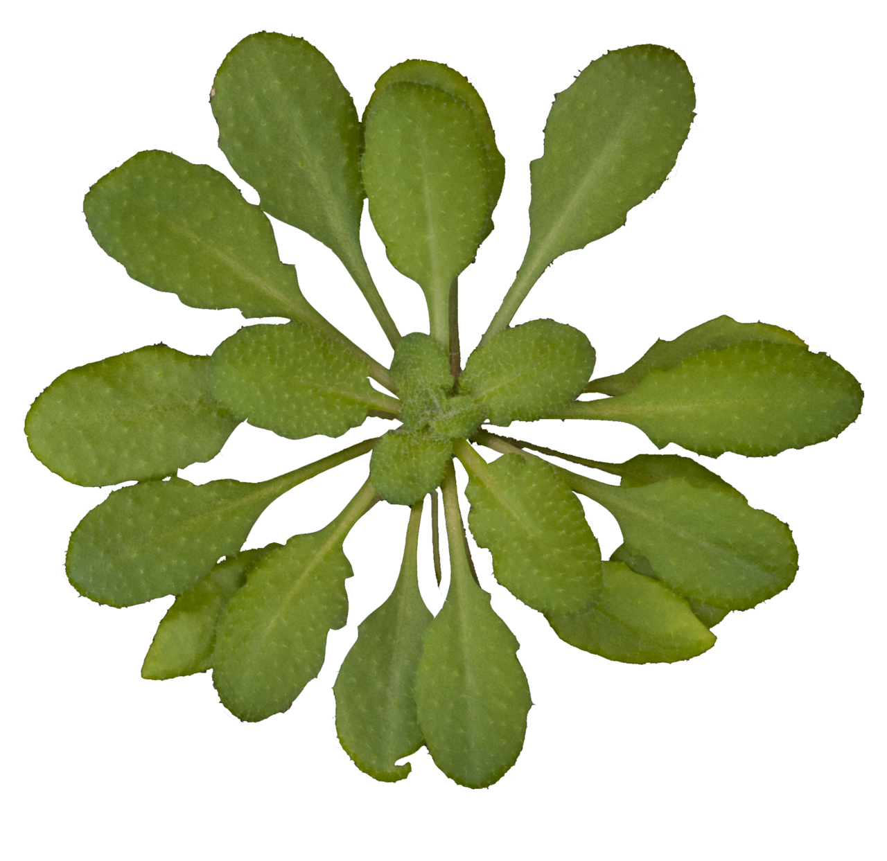
Charles Andrès · Source · CC BY-SA 3.0

The brightest part of a glowing leaf can draw attention away from where the plant is responding to infection. In a new experiment, shining veins dominate one image; a second view reveals a concentrated response around the infection site. Comparing two colors makes an otherwise hard-to-see piece of plant biology visible.
The comparison comes from a study published October 23 in Nature Plants. Viktor Morozov and colleagues developed light-producing reporters—genetically engineered systems whose glow helps researchers follow gene activity. Their advance addresses a deceptively simple problem: a brighter plant does not necessarily mean a more active gene.
You can see the problem in Figure 2e, at the lower right of the published figure. It shows an engineered Arabidopsis thaliana leaf at two times, labeled zero and 150 minutes. The researchers introduced the bacterial pathogen Pseudomonas savastanoi into the leaf; an arrow marks the treated area.
First, look at the upper, black-and-white images. These record light in the green part of the spectrum. At 150 minutes, the veins stand out sharply. Now compare the images underneath: an orange-red patch appears around the marked infection site. That localized response is much easier to distinguish from the branching veins.
The lower images are calculated maps. At each image position, the researchers divided the green-light reading by a reading in a second wavelength band, then assigned the resulting number a display color. The orange and red show differences in that ratio; they are not photographs of the leaf visibly turning those colors. For this infection experiment, the team used a laboratory imager with filters to separate the light bands.
Why should division reveal something that brightness obscures? The plants carry a light-producing system based on fungal bioluminescence. They manufacture the chemical that powers the glow. Its supply depends on the plant’s metabolism—the chemical work that keeps its cells functioning. Oxygen availability and other conditions can also affect light output. A change in brightness therefore has several possible explanations.
The researchers paired two engineered light-producing proteins, called luciferases. Both use the same chemical substrate, but emit different colors. Production of the green protein follows the genetic activity being investigated. A red-emitting protein provides a reference, controlled by a separate DNA sequence intended to remain active. Comparing their outputs reduces interference from changes affecting their shared light-producing chemistry. The paper reports substantially steadier measurements using this approach.
For a simple illustration, imagine a green reading of 100 and a reference reading of 50: the ratio is two. If a shared metabolic change lowers them to 40 and 20, the ratio remains two. Those are illustrative numbers, but they show why a leaf can become much dimmer without implying that the monitored genetic activity has fallen.
In the infection demonstration, the green reporter was controlled by a DNA sequence responsive to jasmonic-acid signaling. Jasmonic acid is a plant hormone. The map therefore follows the activity of a chosen hormone-responsive genetic control sequence; it does not directly measure hormone concentrations. Keeping the location visible matters: a concentrated response around an infection site tells a different story from a change spread evenly across a leaf.
The authors also checked selected reporters using measurements of RNA, the working copies produced when genes are active. In tobacco leaves treated with a growth regulator, changes in the color ratio correlated with changes measured by an RNA assay. That assay was more sensitive at low treatment levels. A separate defense reporter in Arabidopsis also showed agreement between light measurements and gene transcription after chemical stimulation. These were checks of particular reporter systems, distinct from the infection-image demonstration.
The researchers’ institutional announcement, carried by Phys.org, suggests future plants could signal disease or environmental conditions through their glow. Those remain proposed applications rather than demonstrated field monitoring. The paper discloses partial funding by Light Bio and Planta.
Even the reference needs checking. Its controlling DNA can behave differently between tissues, and its activity may depend on where the inserted genes land in the plant’s genome. The authors therefore recommend testing conclusions from the images with other measurement methods. Two colors make the glow easier to interpret; RNA measurements and other checks help establish what that interpretation can support.
Sources:
Sources
Discussion
Kind, curious discussion is welcome. Comments are checked before appearing. Requests to direct the newsroom are discarded.