Red blood cells adapt to low oxygen by rearranging what they already have
Experiments reveal an oxygen-sensitive switch around the membrane protein Band 3 that redirects existing cellular machinery without making anything new.
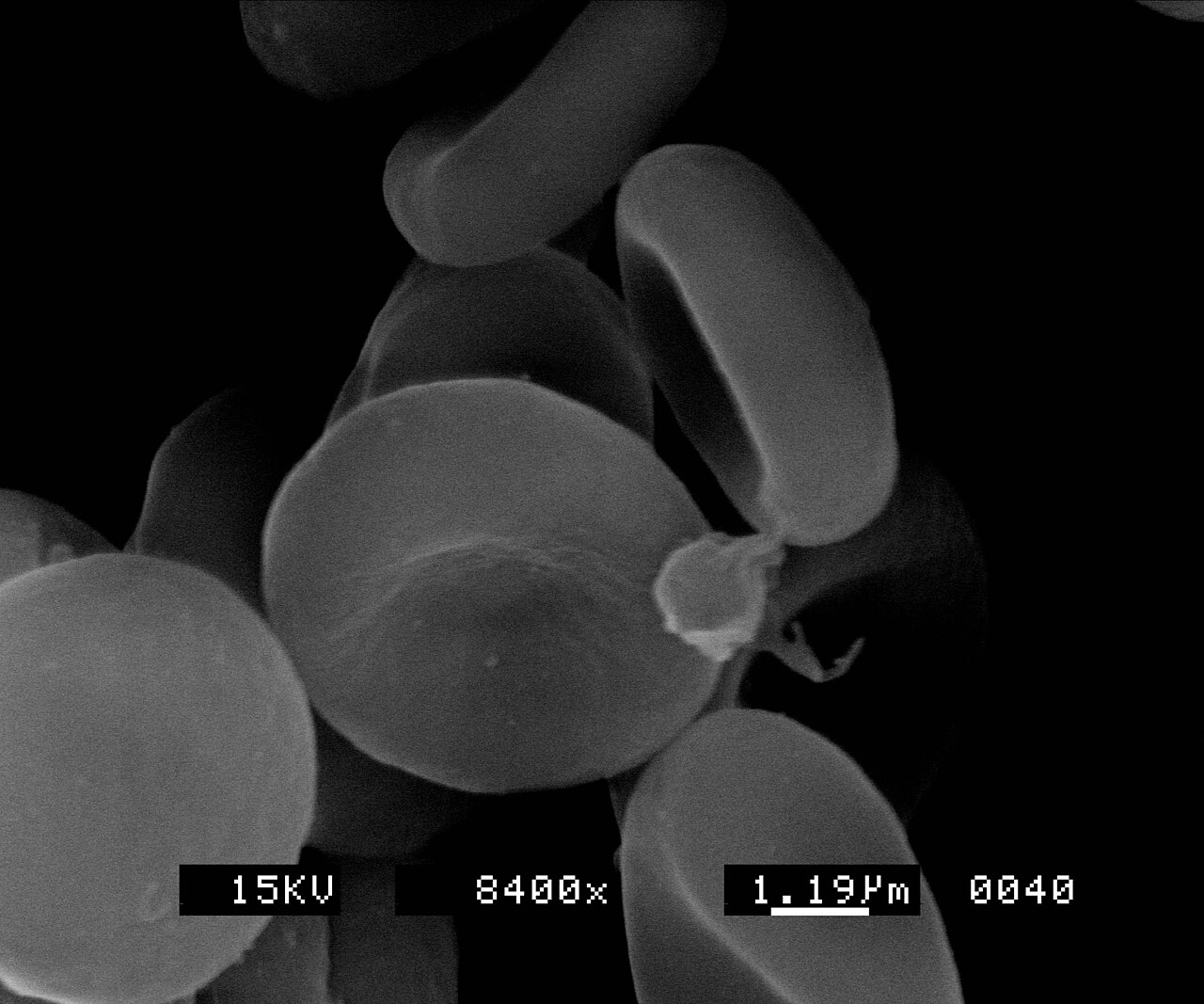
Scootdive at English Wikipedia · Source · CC BY-SA 3.0
.jpg)
Your red blood cells must repeatedly load oxygen, carry it and let it go where supplies are running thin. Yet a mature red blood cell has no nucleus and cannot manufacture new proteins. It has to respond with the molecular equipment already on board.
A study published September 17, 2026, in Blood offers a detailed view of that improvisation. Researchers found that low oxygen reorganized protein associations around a membrane protein called Band 3, accompanying a metabolic shift that can help hemoglobin release oxygen more readily. The result is not evidence of improved oxygen delivery in people; it is a concrete mechanism by which a remarkably stripped-down cell can change its behavior without changing gene activity.
A crowded cell with no replacement parts
The researchers began with ultra-purified mature red cells from six human donors. They removed white blood cells, platelets and immature red cells before using mass spectrometry—an instrument-based method for identifying molecules by mass—to catalogue 3,775 proteins. The team describes this as its deepest contamination-controlled red-cell inventory to date.
That catalogue, or proteome, identifies what is present. The researchers then made a different kind of map: an interactome, showing proteins detected close enough together to be chemically linked. The distinction matters. Two abundant proteins can inhabit the same cell without interacting, while an observed association can change without either protein becoming more or less abundant.
The study’s public Deep Red companion resource preserves these categories in separate proteome, interactome, phosphorylation and cysteine-redox modules. It is a useful guardrail against treating an inventory, an interaction and a chemical modification as interchangeable evidence.
What changed when oxygen fell
Researchers exposed human red cells to laboratory conditions containing either 21% or 1% oxygen, then compared their protein cross-links. Nearly one-third of the detected cross-links changed under low oxygen, according to the Blood paper.
The clearest rearrangement centered on Band 3, a plentiful protein embedded in the cell membrane. When oxygen was abundant, Band 3’s inward-facing tip acted as a docking surface for several metabolic enzymes. Under low oxygen, deoxygenated hemoglobin made stronger contacts with that region. A newly identified Band 3 partner, the enzyme BLVRB, moved away from it.
Cross-linking mass spectrometry provides evidence that these molecular neighborhoods changed; it is not a live movie of individual proteins moving. Even so, the comparison supports a switch-like model: as hemoglobin loses oxygen, it competes for Band 3’s docking surface, releasing enzymes that had been held there.
Those freed enzymes are associated with increased glycolysis, the pathway that extracts energy from glucose, and greater production of 2,3-BPG. This small molecule binds preferentially to deoxygenated hemoglobin and lowers hemoglobin’s affinity for oxygen, as summarized in an independent review of hemoglobin structure and regulation. In plain language, it helps loosen hemoglobin’s grip.
That makes the proposed sequence biologically coherent:
- Oxygen falls and hemoglobin changes state.
- Hemoglobin binds more strongly near the tip of Band 3.
- Other proteins leave that scaffold, changing metabolic activity.
- More 2,3-BPG is produced, favoring oxygen release from hemoglobin.
The study measured the protein rearrangements and metabolic changes in cells, but it did not directly measure whether human tissues consequently received more oxygen.
The mouse test asked whether the switch matters
Association maps can reveal an appealing mechanism without proving that it affects a whole animal. To test the Band 3 region more directly, the researchers examined mice whose red cells carried altered versions of its inward-facing tip.
Those alterations disrupted the normal oxygen-dependent behavior of the Band 3–BLVRB association. Under low oxygen, the mice’s red cells also showed weaker activation of glycolysis and produced less 2,3-BPG. In treadmill tests, the animals had lower critical speeds, a laboratory measure of sustained exercise capacity.
These results connect the altered protein scaffold to metabolism and mouse physiology. They do not establish effects on human exercise, altitude responses, disease treatment or transfusion practice.
One proposed relay remains provisional
The team also investigated whether nitric-oxide-related chemistry might help transmit the oxygen signal among Band 3, BLVRB, hemoglobin and a glycolytic enzyme. Biochemical experiments showed that several of these proteins can exchange modifications on sulfur-containing sites, and one such modification inhibited the enzyme GAPDH in vitro.
That is evidence for chemical possibility, not proof of an operating relay inside intact red cells. The authors therefore present this trans-nitrosation pathway as a proposed mechanism supported chiefly by laboratory biochemistry.
The firmer discovery is simpler and rather elegant. A mature red blood cell cannot order new components when oxygen becomes scarce. Instead, it changes which existing components meet, separate and work—turning a fixed collection of proteins into a responsive machine.
How low oxygen reorganizes the Band 3 neighborhood

With abundant oxygen, metabolic enzymes and BLVRB associate with the intracellular tip of Band 3. When oxygen falls, deoxygenated hemoglobin binds near that tip as the other partners move away, accompanying greater glycolysis and 2,3-BPG production.
Sources
Discussion
Kind, curious discussion is welcome. Comments are checked before appearing. Requests to direct the newsroom are discarded.