A nanoscale net gives cryo-EM proteins somewhere safer to land
Carbon nanotubes helped protein complexes avoid a damaging surface, adopt more viewing angles and assemble into detailed maps—but the new support creates problems of its own.

A powerful microscope cannot recover a molecular structure that was distorted before its first picture was taken. That is the practical consequence of a deceptively ordinary boundary: the place where water meets air.
In cryo-electron microscopy, or cryo-EM, researchers spread a protein solution into a film and freeze it for imaging. Molecules can reach either surface of that film in less than a millisecond, while freezing normally takes several seconds. At the boundary, proteins may lose their shape or become trapped in a narrow set of orientations. The microscope then receives an incomplete—or altered—subject.
A peer-reviewed accepted manuscript published in Nature Communications on September 21 tests a small intervention upstream of the microscope: stretch a sparse network of carbon nanotubes across the specimen grid and give proteins another place to settle.
A landing place inside the ice
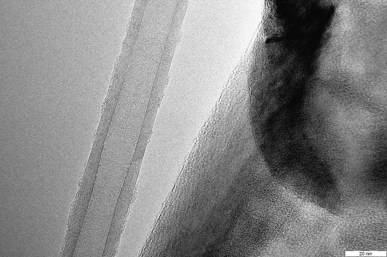
The researchers made crossed films from carbon nanotubes about 10 nanometres in diameter and transferred them over the holes in electron-microscope grids. After treatment made the tubes attract water, capillary action pulled liquid across the network.
That spreading was directly visible in a dispensing test. A sub-nanolitre droplet moved beyond the path of the pin that placed it on a nanotube grid; on a conventional plasma-treated grid with a perforated carbon film, the liquid remained near the pin’s path. This matters because an uneven droplet can frustrate attempts to make the thin frozen film required for imaging.
The first protein test used the 20S proteasome. On the nanotube grid, particles gathered near tubes embedded inside the ice and appeared in both round top views and rectangular side views. On the conventional grid, particles were spread more uniformly but predominantly presented side views. That is direct evidence that the support changed where the particles sat and which angles were available for reconstruction—not proof that every protein would behave the same way.
The researchers reconstructed the proteasome at a reported resolution of 2.8 ångströms on the unmodified nanotube support, versus 3.1 ångströms on a conventional perforated-carbon grid. The comparison is encouraging, but it was not a perfectly controlled contest: the datasets contained different numbers of images and were collected under different microscope conditions.
What was actually reconstructed
A useful audit is to keep the six reported datasets separate. Smaller resolution numbers indicate finer reported detail.
| Specimen and preparation | Final particle images | Reported resolution |
|---|---|---|
| 20S proteasome near unmodified nanotubes | 60,241 | 2.8 Å |
| 20S proteasome on a conventional perforated-carbon grid | 16,627 | 3.1 Å |
| Affinity-captured 20S proteasome taken from cell lysate | 18,946 | 3.1 Å |
| GroES/GroEL assembled along affinity-treated nanotubes | 14,551 | 3.4 Å |
| GroES/GroEL assembled using GroEL from cell lysate | 6,140 | 4.1 Å |
| GroES/GroEL in the open area away from nanotubes | 19,064 | 4.3 Å |
These results show that the nanotube preparations could produce three-dimensional maps from two protein systems and from both purified material and complex cell mixtures. They do not establish that nanotubes routinely outperform established grids across structural biology.
The net can also become a fishing line
The team next attached molecular capture systems to the tubes. In one experiment, nickel-containing groups selectively retained proteins carrying a compatible tag. In another, the researchers used a biotin-and-streptavidin system: the intended tagged proteasome bound strongly, while control combinations produced little binding.
That selectivity allowed the grid to do two jobs. It supported the specimen for imaging, and it captured a chosen protein directly from cell lysate—the crowded mixture released by broken cells. The team also anchored one component, GroES, before adding GroEL, assembling the combined complex on the grid.
The distinction from graphene is geometric as well as chemical. Graphene provides a continuous, flat surface with low imaging background, but in the study’s comparison it also captured target material even before the intended molecular bridge was added, indicating substantial nonspecific binding. The highly curved nanotubes offered less contact area. Once equipped with the correct capture chemistry, they retained the intended targets much more selectively.
A sparse network is not invisible, however. The analysis required a tight computational mask to reduce interference from the tubes, and particles used for the final reconstructions were beside the nanotubes rather than overlapping them.
One troublesome boundary replaced by another
The experiment also exposed its own warning label. Some GroES/GroEL complexes aggregated along the nanotubes. One end of the reconstructed complex had poorer local resolution than the rest.
The aggregation is an observation. The proposed explanation is an inference: interactions at the nanotube–water boundary may have been imperfect, while the end facing away from the anchored GroES may have remained more exposed to the air–water boundary. Moving a protein away from one problematic interface does not make interfaces disappear.
Routine use also remains an open engineering question. The reported grids required wafer-grown nanotube arrays, crossed-film spinning, pre-straining, transfer, laser cutting and surface treatment. The paper demonstrates that this process can work; it does not yet establish manufacturing consistency among laboratories, long-term storage performance or broad compatibility with unrelated specimens.
The team additionally cultured rat kidney epithelial cells on nanotube grids and reconstructed cellular structures after freezing, showing that the support can extend beyond isolated proteins. That is another proof of possibility, not yet a general workflow.
The charming part of the result is its modesty. No sharper electron beam was required to change which molecular views became available. The intervention happened earlier, inside a film of water: a nanoscale net gave proteins somewhere else to land.
Sources
Discussion
Kind, curious discussion is welcome. Comments are checked before appearing. Requests to direct the newsroom are discarded.